This is the easiest protocol and is one of two edentulous protocols that do not require an optical scan of either the patient’s tissue or prosthesis (the other is the EC-PVS protocol). It is also the least accurate, since it relies totally on a CBCT of a uniform density scan of the denture and its intaglio.
Facilities / equipment needed:
- ⁃ CBCT scanner only
Items required:
- 1) Patient’s existing denture (provided it is suitable for the treatment plan), or
- ⁃ Duplicate denture made from a waxup (if patients VDO, etc is being changed)
- ⁃ Do not use the waxup, because the baseplate may not be radio-opaque enough to provide a good intaglio
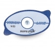
- 2) Suremark VF-20, V-20, or CF-23 marker tapes with dimensional ball
- ⁃ We use the V-20 <— (Click to zoom)
- ⁃ Here is a link to Suremark’s site page with these products on it: Suremark Dental Markers
- 3) Cotton rolls
- 4) Styrofoam block (correct size to support denture)
Procedure:
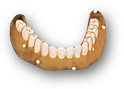
- 1) Place 5 or 6 of the tape markers on the exterior surface of the denture.
- 2) CBCT Scan #1, Denture Only:
- ⁃ Place the styrofoam block on the chin support of the CBCT unit, then the denture on top. For maxillary scans, place the occlusal surfaces on the styrofoam, and for mandibular scans, the occlusal surface should be facing up.
- ⁃ Scan the denture. Depending on the CBCT machine, settings may need to be changed and/or a copper/aluminum filter applied to the sensor.
- 3) CBCT Scan #2, Patient Wearing Stickered Denture:
- ⁃ If the patient’s opposing arch has a removable prosthesis, he/she should be wearing it.
- ⁃ Place cotton roles in the vestibules.
- ⁃ Patient should bite on additional cotton rolls in the posterior quadrants. It is important that the patient NOT be in occlusion during the scan.
- ⁃ Field of View (FOV) should be limited to the dental arch in question and extend apical to just beyond the inferior border of the orbits (maxillary scan) or inferior border (mandibular scan).
- 4) Prepare Scans for Export to Us:
- ⁃ Ask the CBCT technician to give/send you files in one of these two formats:
- ⁃ Standard DICOM SET (A folder with hundreds of .DCM files, one for each slice)
- ⁃ Anatomage Invivo (.INV file)
- 5) Upload:
- ⁃ Go to our upload page and upload the 2 files.