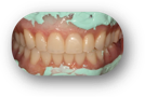
This is an improved, more accurate CBCT-only approach and is one of two edentulous protocols that do not require an optical scan of either the patient’s tissue or prosthesis (the other is the EC-D protocol). It was developed by Dr. Michael Scherer (whose photography is used above, with permission) and it provides a vastly improved CBCT image of the intaglio of the denture; the best possible without utilizing an optical scan. You can view his description of this technique in his Linkedin article: The role of radiopaque PVS in CBCT imaging for full-arch reconstruction.
Finally, unlike the EC-D protocol, you can use an unprocessed denture waxup, since the intaglio is captured with great accuracy. This saves the time and cost of making a duplicate appliance.
Facilities / equipment needed:
- ⁃ CBCT scanner only
Items required:
- 1) Patient’s existing denture (provided it is suitable for the treatment plan), or
- ⁃ If the treatment plan departs from the setup of the existing denture, a waxup is just fine.
- 2) Parkell Green-Mousse (NOT Blue-Mousse), SKU: S455S <— (Click to zoom)
- 3) OPTIONAL: Suremark dental markers.
- ⁃ This helps in aligning the denture scan to the patient scan.
- ⁃ Here is a link to Suremark’s site page with these products on it: Suremark Dental Markers. We use the V-20 <— (Click to zoom)
- 4) Cotton rolls.
- 5) Styrofoam block (correct size to support denture).
Procedure:
- 1) (OPTIONAL) place 2 or 3 Suremark stickers on the denture. This helps us more efficiently merge the denture scan into the patient scan.
- 2) With the patient seated and opposing arch in place, apply the Green Mousse to the intaglio, insert in patient’s mouth, and ask the patient to occlude normally. No adhesive is necessary.
- 3) CBCT Scan #1, Patient Wearing PVS Relined Denture:
- ⁃ If the patient’s opposing arch has a removable prosthesis, he/she should be wearing it.
- ⁃ Patient should bite on cotton rolls in the posterior quadrants. It is important that the patient NOT be in occlusion during the scan.
- ⁃ Field of View (FOV) should be limited to the dental arch in question and extend apical to just beyond the inferior border of the orbits (maxillary scan) or inferior border (mandibular scan).
- 4) CBCT Scan #2, Denture Only:
- ⁃ Place the styrofoam block on the chin support of the CBCT unit, then the denture on top. For maxillary scans, place the occlusal surfaces on the styrofoam, and for mandibular scans, the occlusal surface should be facing up.
- ⁃ Scan the denture. Depending on the CBCT machine, settings may need to be changed and/or a copper/aluminum filter applied to the sensor.
- 5) Prepare Scans for Export to Us:
- ⁃ Ask the CBCT technician to give/send you files in one of these two formats:
- ⁃ Standard DICOM SET (A folder with hundreds of .DCM files, one for each slice)
- ⁃ Anatomage Invivo (.INV file)
- 6) Upload:
- ⁃ Go to our upload page and upload the 3 files.